


Jakob Bratkovič: Postcard from Madison
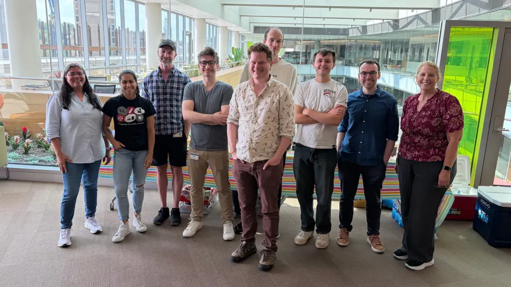
Exploring Medical Imaging, Brain Biomarkers, and Optical Microscopy
Written by: Jakob Bratkovič
It has been a great opportunity to spend my research visit in Madison, Wisconsin, where I worked in an interdisciplinary research environment at the University of Wisconsin–Madison. The visit was made possible through the invitation of Professor Robert Jeraj and allowed me to gain experience in two complementary areas: medical image analysis and optical microscopy.
A major part of my work focused on brain imaging and quantitative image analysis. I worked with SPM templates and explored the role of anatomical templates in spatial normalisation and the comparison of brain images across different subjects. This helped me better understand how preprocessing choices can influence later quantitative analyses.
I also worked extensively with FreeSurfer and FastSurfer, two tools used for automated brain segmentation and anatomical analysis. Through these pipelines, I gained practical experience with the segmentation of cortical and subcortical structures and with extracting quantitative measurements from different brain regions.
A significant part of the project focused on the statistical analysis of these measurements. I analysed volumes of cortical and subcortical structures and investigated variability, relationships between anatomical regions, and differences between subjects. One of the main goals was to explore whether these measurements could be useful for identifying potential imaging biomarkers.
I was particularly interested in whether combinations of volumetric measurements could reveal patterns that may not be apparent when individual regions are considered separately. This part of the work gave me more experience with statistical analysis and with interpreting quantitative results derived from medical images.
Another important part of my project was brain asymmetry. I investigated differences between corresponding structures in the left and right cerebral hemispheres and explored how these differences could be quantified from automated segmentations. I looked at both cortical and subcortical structures and considered whether asymmetry measures could provide additional information beyond absolute volume measurements.
This also required careful evaluation of the reliability of the results, since observed differences may reflect genuine anatomy but can also be influenced by image acquisition, preprocessing, segmentation methods, and natural variability between individuals. This made it important to consider the entire image-processing pipeline when interpreting the findings.
During this part of the fellowship, I had the opportunity to collaborate with Alison Deatsch on the analysis and interpretation of brain imaging data. Our discussions were useful for understanding the methodological aspects of the analysis and for putting the quantitative results into a broader context.
In addition to computational medical imaging, I gained hands-on experience in optical microscopy in Randy Bartels’ laboratory, where I worked under the guidance of Matthew Sheinman. This part of the visit offered a different perspective on imaging, as I was able to work directly with optical systems and learn more about the physical principles behind image formation.
As part of this work, I used phase and polarisation-sensitive cameras, which can provide information beyond conventional intensity imaging. Working with these techniques helped me better understand how phase and polarisation can reveal properties of a sample that are not as easily observed with standard imaging methods. I also gained practical experience with optical setups, image acquisition, and the interpretation of experimental data.
As part of the experimental work, I prepared and imaged samples containing polystyrene beads and TiO₂ particles. These controlled samples were useful for testing the imaging system and comparing results obtained under different conditions. Through this work, I gained experience with sample preparation, microscope alignment, image acquisition, and experimental image analysis.
Another part of the microscopy project involved image stitching. Since the field of view of a microscope is limited, larger areas can be reconstructed from several partially overlapping images. I worked on aligning neighbouring images and combining them into larger mosaics.
This introduced me to several practical challenges that come with real experimental data, including variations in illumination, small changes in sample position, imperfect overlap between images, and the need for accurate registration. It was also a useful way to apply image-processing concepts in a new experimental setting.
Working under the guidance of Matthew Sheinman gave me the opportunity to discuss both the theoretical and practical aspects of the experiments. I gained a better understanding of how different parts of the optical system contribute to the final image and how choices made during an experiment affect the information that can later be extracted from the data.
Overall, the fellowship allowed me to work with two different but closely related approaches to imaging. On one side, I worked with computational analysis of medical images of the brain, focusing on statistical analysis, structural volumes, asymmetry, and potential imaging biomarkers. On the other, I gained direct experimental experience with microscopy, phase and polarisation imaging, sample preparation, and image stitching.
Through this work, I developed my skills in medical image processing, brain segmentation, statistical analysis, biomarker exploration, optical microscopy, image acquisition, image stitching, and scientific interpretation of data. I also gained a better sense of how different disciplines can contribute to the same broader goal of extracting useful information from biomedical images.
Beyond the research, I greatly enjoyed living in Madison and experiencing the atmosphere of an American university city. One of the highlights of my stay was celebrating the Fourth of July, which gave me the chance to experience an important American tradition first-hand and made the stay even more memorable.
During my time in the United States, I also had the opportunity to visit Oklahoma and Washington, D.C.. Seeing different parts of the country added an important cultural aspect to the fellowship and gave me a broader experience of life in the United States beyond Madison.
Working with researchers from different backgrounds was also an important part of the visit. I appreciated the opportunity to ask questions, exchange ideas, learn about different research approaches, and build professional connections. We plan to stay in contact and continue collaborating on projects beyond the fellowship, which is one of the outcomes of the visit that I value most.
I am especially grateful to Professor Robert Jeraj for the invitation and for making this research visit possible, as well as to Alison Deatsch, Matthew Sheinman, and the wider research teams for their guidance, discussions, and support during my stay. I am also sincerely grateful to the ASEF team for making this research and cultural experience possible.
My time in Madison broadened my understanding of biomedical imaging and gave me valuable research experience, new technical skills, and professional connections that I hope to continue developing in the future.